Exercise has profound and positive effects on the aging population, improving physical, mental, and social well-being. Here are some key benefits:
You
- Physical Benefits
Cardiovascular Health
How it helps: Aging naturally weakens the heart and blood vessels, leading to increased risks of heart disease. Regular aerobic exercise like brisk walking, swimming, or dancing strengthens the heart muscle, improves circulation, and lowers bad cholesterol (LDL) levels while boosting good cholesterol (HDL).
Result: Reduced risk of hypertension, heart attacks, and strokes.
Musculoskeletal Health
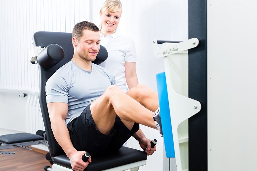
How it helps: After age 30, muscle mass begins to decline (sarcopenia). Strength training with light weights or resistance bands helps maintain and build muscle mass. Weight-bearing exercises like walking or jogging help strengthen bones, reducing the risk of osteoporosis.
Result: Improved strength, posture, and joint stability.
Mobility and Flexibility
How it helps: Aging often leads to stiffness and reduced range of motion. Stretching exercises and activities like yoga improve flexibility, while balance exercises (e.g., tai chi) reduce the risk of falls and injuries.
Result: Enhanced independence and reduced need for assistance in daily activities.
Immune Function
How it helps: Regular moderate-intensity exercise boosts the production of immune cells and enhances their activity.
Result: Older adults can better resist infections and recover faster from illnesses.
- Cognitive Benefits
Slowing Cognitive Decline
How it helps: Exercise increases blood flow to the brain and stimulates neurogenesis (the growth of new neurons), which helps preserve cognitive function.
Result: Lower risk of dementia, Alzheimer’s, and age-related memory loss.
Enhancing Memory and Learning
How it helps: Physical activity promotes the release of brain-derived neurotrophic factor (BDNF), which supports brain plasticity (the brain’s ability to adapt and change).
Result: Better problem-solving skills, memory retention, and decision-making.
Reducing Stress and Anxiety
How it helps: Exercise triggers the release of endorphins, serotonin, and dopamine—natural mood elevators. It also reduces levels of cortisol, a stress hormone.
Result: Improved mood, reduced risk of depression, and better coping mechanisms.
- Managing Chronic Diseases
Diabetes
How it helps: Exercise improves insulin sensitivity, allowing the body to use blood glucose more effectively. Activities like walking after meals can help stabilize blood sugar levels.
Result: Better management of Type 2 diabetes and reduced risk of complications.
Hypertension and High Cholesterol
How it helps: Regular physical activity strengthens the heart, which lowers blood pressure and improves blood flow. It also promotes the breakdown of fats in the bloodstream.
Result: Reduced risk of cardiovascular events.
Arthritis
How it helps: Low-impact exercises like swimming or water aerobics reduce stress on joints, while strength training supports joint stability.
Result: Decreased pain, better mobility, and improved quality of life.
- Social and Emotional Benefits
Social Interaction
How it helps: Group exercise settings foster connections with peers, which is particularly important for older adults who may face isolation.
Result: Reduced feelings of loneliness and increased emotional well-being.
Enhanced Quality of Life
How it helps: Regular activity improves energy levels, sleep patterns, and self-confidence, enabling older adults to engage more fully in life.
Result: Greater satisfaction and independence in daily living.
- Longevity
How it helps: Studies consistently show that active individuals live longer than their sedentary counterparts. Exercise reduces the risk of chronic diseases and promotes overall body resilience.
Result: Increased life expectancy and healthier years of life.
Recommended Types of Exercise
- Aerobic Activities:
Examples: Brisk walking, cycling, swimming, or dancing.
Benefits: Boosts heart health, endurance, and overall energy.
- Strength Training:
Examples: Using light weights, resistance bands, or bodyweight exercises like squats or push-ups.
Benefits: Builds muscle mass, supports bone health, and improves balance.
- Flexibility and Stretching:
Examples: Yoga, pilates, or basic stretches.
Benefits: Enhances range of motion and reduces stiffness.
- Balance Exercises:
Examples: Tai chi, standing on one leg, or balance-specific exercises.
Benefits: Reduces fall risks, a leading cause of injury among older adults.
- Low-impact Activities:
Examples: Water aerobics, chair yoga, or walking.
Benefits: Ideal for individuals with joint pain or mobility issues.
Important Considerations
Start Gradually: Older adults who are new to exercise should begin with low-intensity activities and gradually increase intensity.
Customize for Health Conditions: Exercise programs should be tailored to accommodate chronic conditions like arthritis, heart disease, or diabetes.
Consult a Professional: Always seek advice from a doctor or physical therapist before starting a new fitness regimen.
By integrating regular exercise into their lifestyle, aging adults can significantly enhance their physical, mental, and emotional health, leading to a more active and fulfilling life.